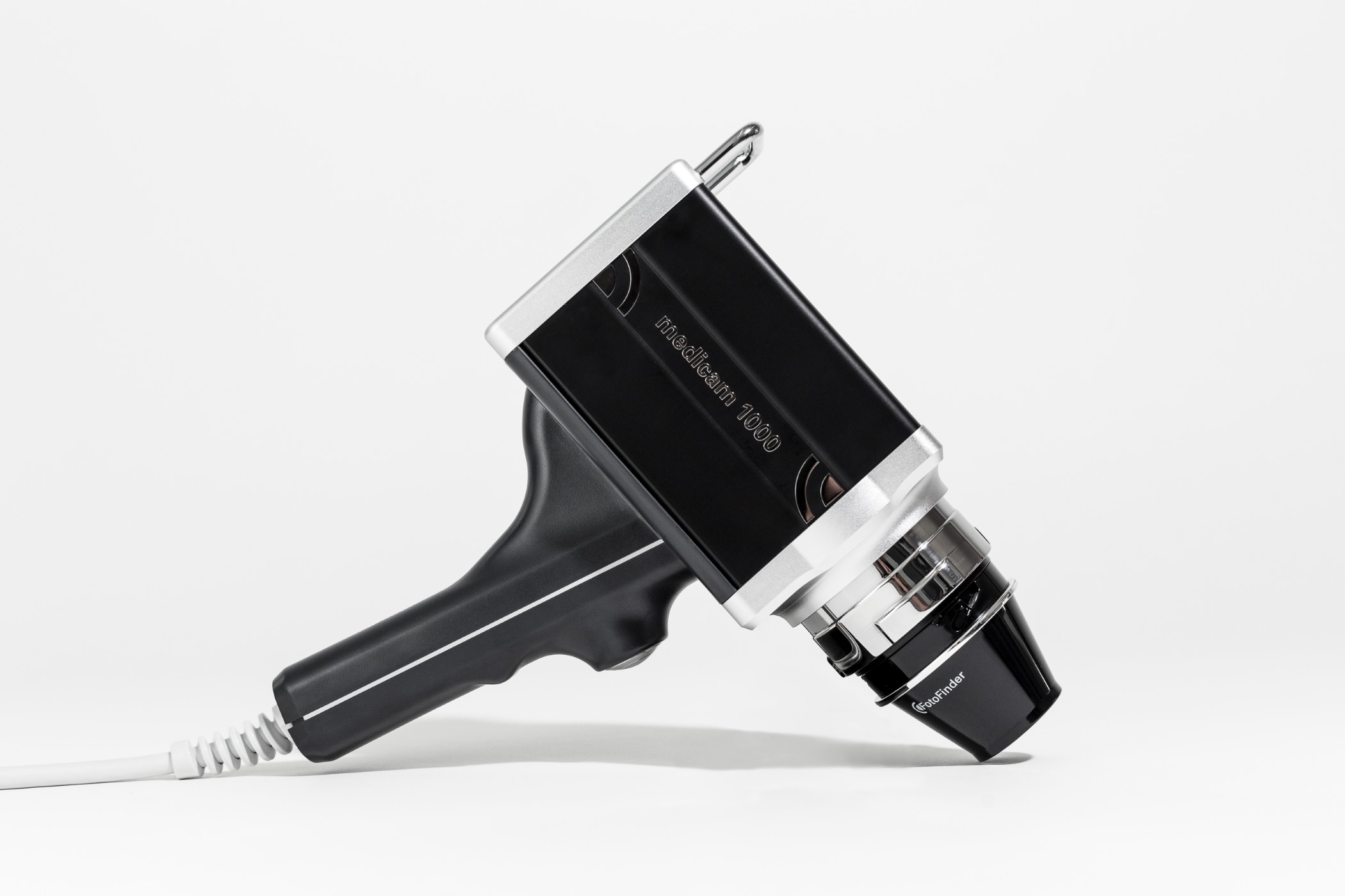
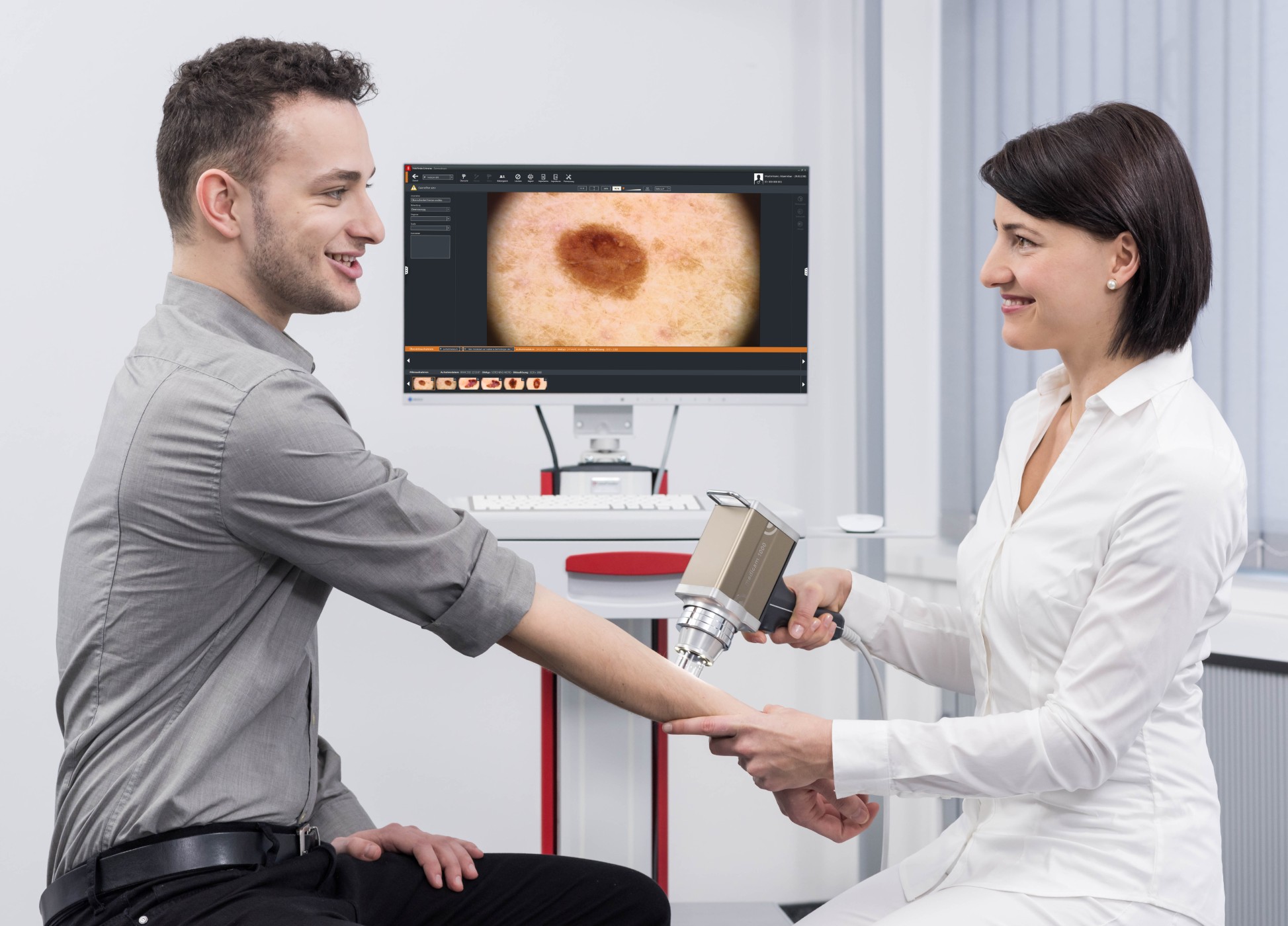
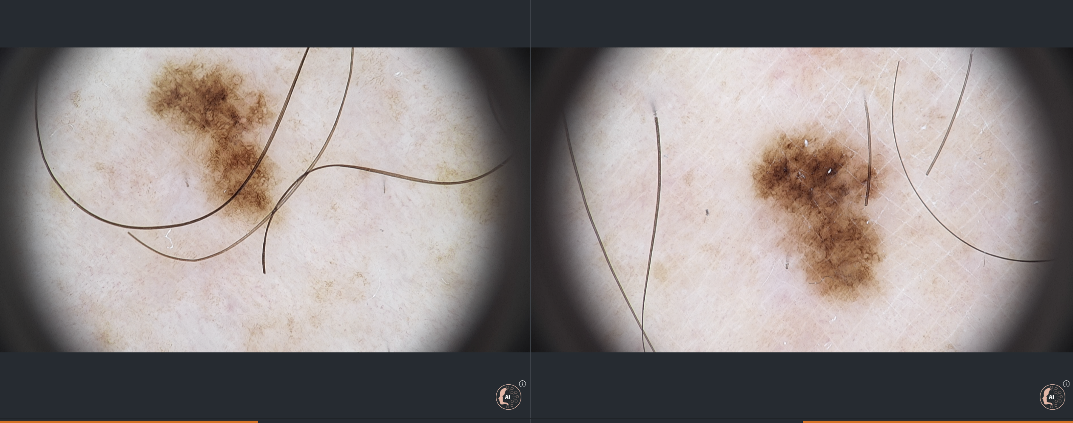
High-Powered Video Dermoscopy and Structural Analysis
With FotoFinder’s video dermoscope, individual lesions are examined using magnified, polarised, high-definition imaging. This reveals fine diagnostic features — including pigment networks, vascular patterns, follicular openings, and dermoscopic structures — that are not visible to the naked eye. The system’s integrated software applies AI-assisted pattern recognition to support risk stratification, helping identify lesions that display atypical morphology or early malignant characteristics.
")
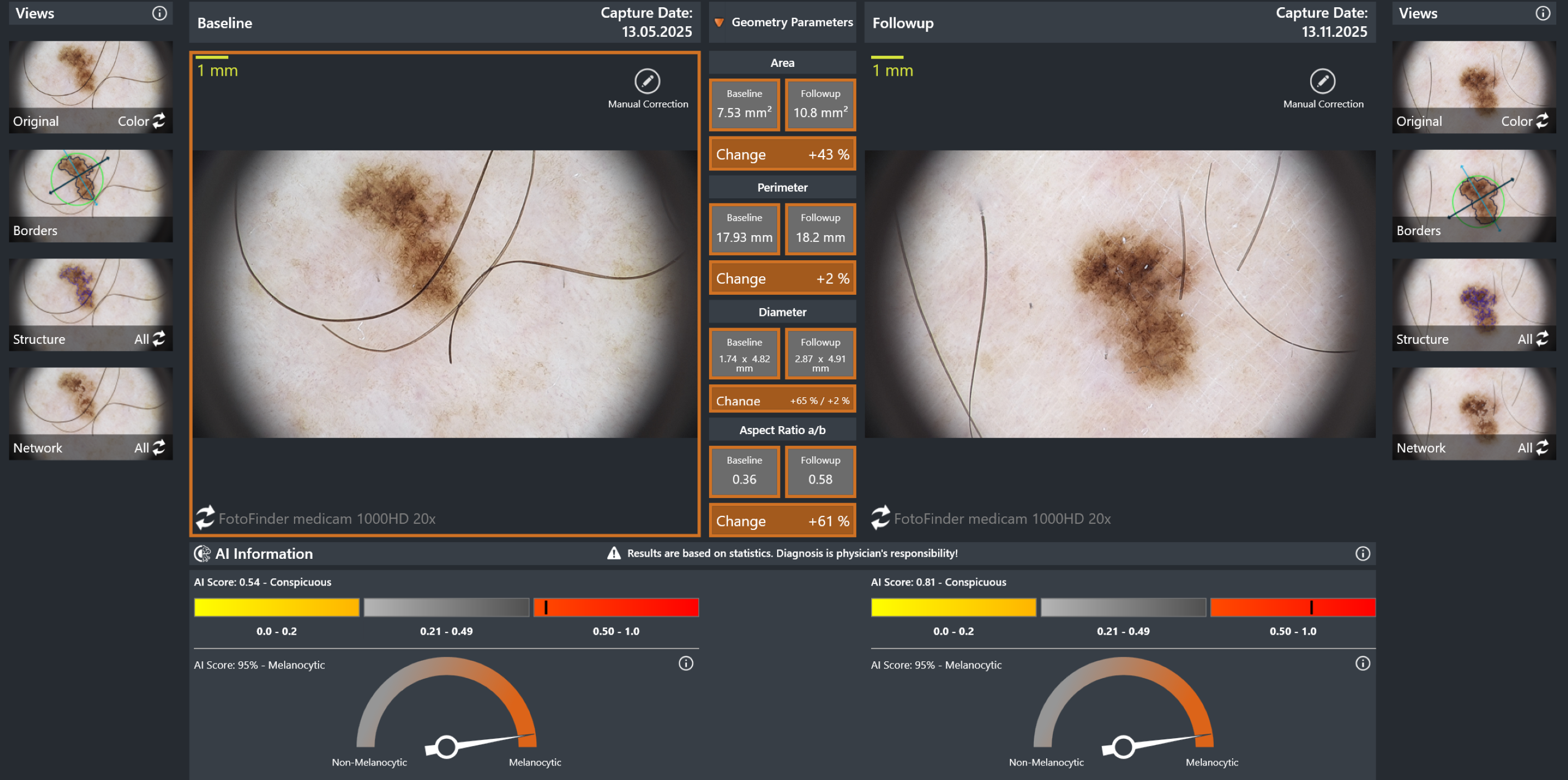
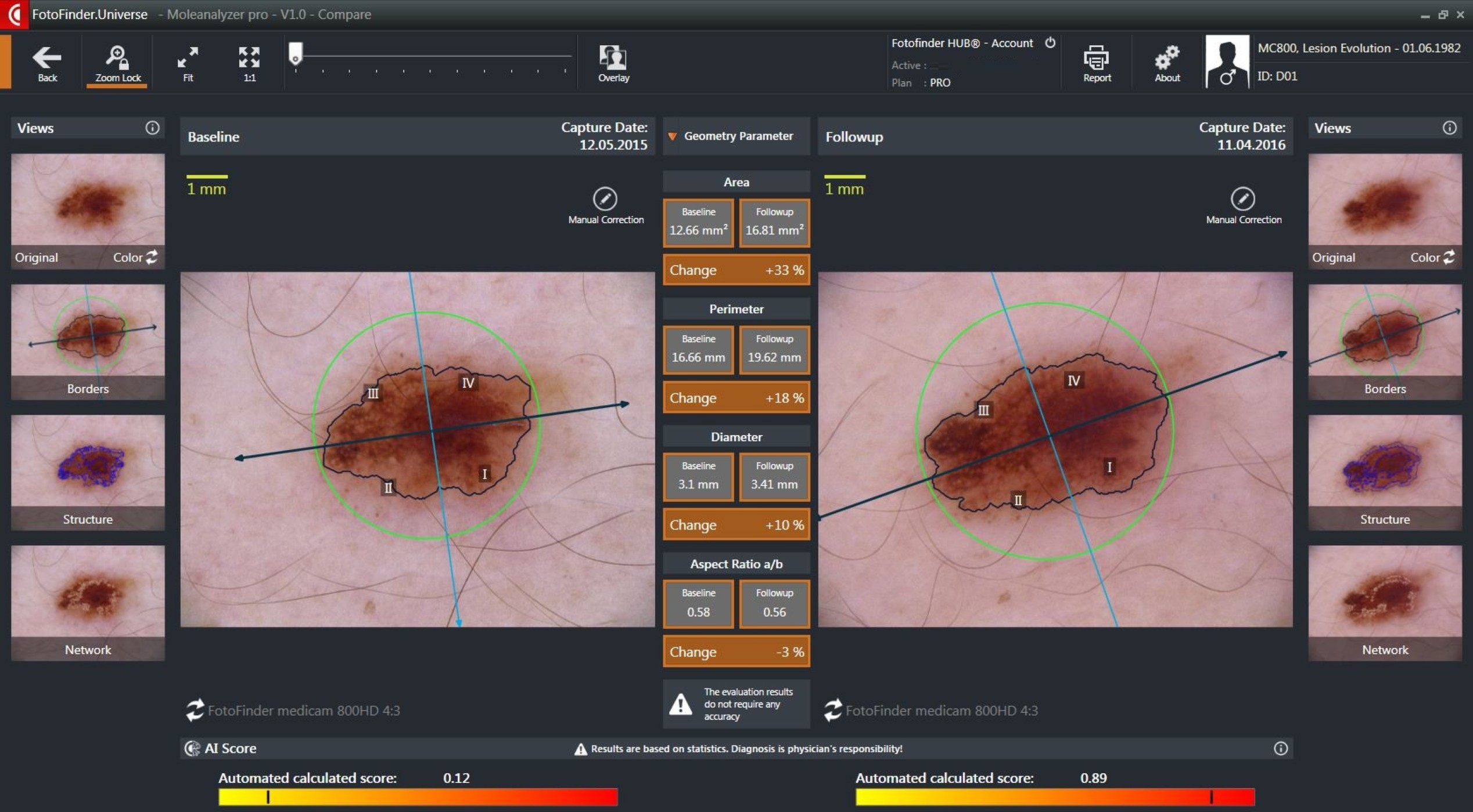
Precision Change Detection Through Longitudinal Monitoring
FotoFinder’s longitudinal imaging software compares current dermoscopic and clinical photos with prior examinations at a pixel-by-pixel level, enabling highly sensitive detection of morphological changes. Even minimal variations in colour, structure, or symmetry — often imperceptible in standard clinical exams — are identified and flagged. This advanced change-mapping significantly enhances early melanoma detection by capturing evolution at its earliest stages, when treatment outcomes are most favourable.